27 Mayıs 2014 Salı

PRINCIPLES OF SKIN COVER - GRAFTS AND FLAPS

Characteristics
  • Largest organ of the human body- surface area 1.5-2.0 sq.m,  2-3mm thick
  • 15% of body weight
  • Epidermis- Corneum, Lucidum, Granulosum, Spinosum, Basale,
  • Dermis- papillary and reticular, made of collagen, 
Functions of skin
  • Protective layer - Langhan’s cells of adaptive immune system
  • Sensation
  • Heat regulation
  • Control evaporation
  • Aesthetics 
  • Makes vitamins D
When is skin required?
  • Loss of large area of skin
  • Large wound 
  • Post burn raw area
  • Release of contracture
Reconstructive Ladder
Graft and Flap
  • Graft is free tissue without its vascular supply- skin, bone, tendon, nerve, vessels, muscle, fascia, mucosa
  • Flap is tissue with its original blood supply
  • Areas where we cannot put graft will need a flap-Flaps for bone devoid of periosteum, cartilage devoid of perichondrium, tendon devoid of paratenon
Grafting 
  • Skin graft is essentially dead with no circulation. Under favourable conditions obtain new blood supply from recepient wound or defect. This is known as graft take
  • Infection, pressure, hematoma or shear forces can result in graft failure
  • Meshing – increase surface area and prevention of collections
  • Immobilisation with Sutures / plaster slab
  • Prevention of hematoma / seroma
  • Dressing of recipient wound
  • Donor site management
Process of graft take

1. Imbibition
2. Inosculation
3. Vascularization

Conditions affecting graft take
  • Malnutrition- hypoproteinemia, vitamin deficiency
  • Presence of hypertension, diabetes
  • Compromised immune status
  • Collagen vascular disease
  • Constant pressure on the area

When to graft?
  • Wound flat and red / can see the imprint of gauge piece
  • No unhealthy granulation tissue or necrotic tissue
  • Margins are healing and spreading
  • Not much bleeding on touch
  • dressing peels  with difficulty
  • Bacterial count less than 105 
  • No evidence of beta hemolytic streptococci


Types of skin graft:


Partial thickness- thin intermediate thick
Full thickness
Composite graft- More than one tissue type required to perform reconstruction
Split thickness skin grafting (Thiersch graft)
  • Donor site heals spontaneously as part of dermis is left behind
  • Contracture may occur
  • Sensation function of hair sebaceous gland not present
  • Donor area is dressed and dressing is removed after 10 days and after 5 days in recepient area

Skin graft harvesting

Instruments  -  Humby’s knife
Donor sites:
Thigh , legs, arm, forearm, body

Taking split skin graft with a Humby’s knife holder

Differences

Split thickness Skin graft:
  • Epidermis and part of dermis
  • Donor sites heal spontaneously 
  • Extensive defects
  • Graft take – good
  • More contraction later
  • Less stable
  • Less colour and texture matching


Full thickness graft:
  • Epidermis and whole dermis
  • Donor site needs to be closed – direct or SSG
  • Small defects
  • Graft take – less readily- needs optimal condition
  • Secondary contraction less
  • More stable
  • Good colour and texture matching


Full thickness skin grafting (Wolfe’s graft)
  • Harvested with surgical blade
  • Accurately fitted to the defect and sutured
  • Donor sites – postauricular, upper eyelid, supraclavicular, flexural, abdomen
  • Donor site closure
  • Recipient site – dressing / immobilisation
  • Whole thickness of skin is excised
  • Defatting is done to improve graft take
  • Blood supply must be restablished
  • Where cosmesis is important (face) or flexibility is important (over joint)

Flaps
  • Flap is tissue with its original blood supply

Classification:

1. Based on blood supply – 

  • Random pattern (dermal and subdermal plexus)

Rotation flap / transposition flap / advancement flap

  • Axial pattern (named vessel) e.g. groin flap (lat. Circumflex artery)

Peninsular flap / island / free flap


Free tissue transfer
  • Most technically demanding
  • Single stage wound closure
  • Wide variety of flaps- tailored to coverage needs
  • More acceptable aesthetic outcome


2. Classification based on tissue type
1. Cutaneous flap
2.Faciocutaneous flap
3.Muscle flap
4.Musculocutaneous flap
5.Osteocuaneous flap

3. Specialized flaps
1. Fascial flap
2. Vascularized bone flap
3. Functional muscle flap

16 Mayıs 2014 Cuma

Signs and Symptoms Associated with Down Syndrome


Down syndrome is the most common autosomal trisomy identified in liveborn infants. As many as 95% of Down syndrome cases arise due to chromosomal nondisjunction during maternal meiosis (47 XX, +21) an abnormality that positively correlates with increasing maternal age. Two of the more prominent and consistent lectures of Down syndrome are mental retardation and facial dysmorphism. Almost every organ and system, however, is affected

Signs and Symptoms Associated with Down Syndrome


Trisomy 21 (Down syndrome) is characterized by mental retardation, facial dysmorphism, single palmar crease, endocardial cushion defects, and duodenal atresia. Affected individuals have an increased risk of AML-M7 and ALL in childhood and early Alzheimer disease in adulthood. 

21 Nisan 2014 Pazartesi

Difference between kawasaki and scarlet fever

Kawasaki disease include: (mucocutaneous lymph node syndrome)


Definition : Kawasaki disease is a vasculitis of medium-sized arteries that affects very young children (usually 12-24 months of age).

cause is unknown

Features:
1. Fever more than 5 days - even after aspirin
2. Bilateral conjunctivitis
3. Lymphadenopathy
4. Cutaneous involvement
1. Oropharyngeal: Erythema of the palatine mucosa, fissured erythematous lips, “strawberry” tongue
2. Peripheral extremities: Edema of hands and feet, erythema of palms and soles, desquamation of the fingertips (periungual)
3. Generalized rash: Polymorphous (usually urticarial) erythematous rash beginning on the extremities and moving to the trunk (centri petal spread)

Complication: Formation of coronary artery aneurysms is the most serious complication of Kawasaki disease.

Scarlet fever : 

Causes : Streptococcus pyogenes (group A streptococci) --Toxigenic

Features:

scarlet rash with Sand paper like texture ,circumoral pallor.


Tonsillar exudates, Spares the palms and soles

Complication: Septic complications

Aspirin helps to reduce fever , Amoxicillin and throat culture

Similarity : ''Strawberry tongue''

29 Mart 2014 Cumartesi

The Gallbladder and bile ducts

Gallbladder
  • The gallbladder is a pear-shaped reservoir in continuity with the common hepatic and common bile ducts through the cystic duct.
  •  It is usually 7.5 to 12cm in length, is 3 to 5 cm in diameter, and has a capacity of 35 to 50 mL.
  •  The gallbladder lies on the inferior surface of the liver partially enveloped in a layer of peritoneum.
  •  The gallbladder is anatomically divided into the fundus, body, infundibulum, and neck, which empties into the cystic duct.
  •  Both the gallbladder neck and the cystic duct contain spirally oriented mucosal folds known as the valves of Heister. 
  • The valves prevent the passage of gallstones and excessive distention or collapse of the cystic duct, despite variations in ductal pressure. 


The Hepatic Duct (ductus hepaticus)

  • Two main trunks of nearly equal size. Arising from the liver at the porta. one from the right, the other from the left lobe.
  • The common hepatic duct is less than 2.5cm long  and is formed by the union of the right  and left hepatic ducts.
  • It passes downward and to the right for about 4 cm.
  • Joined at an acute angle by the cystic duct to form the common bile duct. Lies between  the layers of the lesser omentum.
  • Is accompanied by the hepatic artery and portal vein. 
Cystic duct

The cystic duct varies in length from 1 to 5 cm and in diameter from 1 to 3 mm; it usually joins the common hepatic duct at an acute angle.
The Common Bile Duct 

The common bile duct is formed by the junction of the cystic and hepatic ducts.
  • Length: It is about 7.5 cm. long.
  • Diameter: That of a  goose-quill.


  • Course and extent : It descends along the right free margin  of the lesser omentum along with   portal vein ( which is behind it ) , and to the right of the hepatic artery. 
  • Then behind the superior portion of the duodenum,
  • After crossing the duodenum, it runs on the posterior surface of the head of the pancreas, and in front of IVC. 
  • Occasionally completely imbedded in the pancreatic substance. 
  • At its termination it lies for a short distance along the right side of the terminal part of the pancreatic duct and passes with it obliquely between the mucous and muscular coats. 
  • The two ducts unite and open by a common orifice upon the summit of the duodenal papilla, situated at the medial side of the descending portion of the duodenum, a little below its middle and about 7 to 10 cm. from the pylorus. The short tube formed by the union of the two ducts is dilated into an ampulla, the ampulla of Vater. 
Blood supply
  • Gall bladder is supplied by Cystic artery, a branch of right hepatic artery.
  • The blood supply to the common hepatic duct, cystic duct and common bile duct comes from the gastroduodenal, retroduodenal, postero-superior pacreatico duodenal arteries. 
Lymphatics
  • The lymphatics of gallbladder (subserosal and submucosal) drain into the cystic L/N of Lund.
  • Small veins and lymphatics course between the gallbladder fossa and the gallbladder wall, connecting the lymphatic and venous drainage of the liver and gallbladder. These connections are the cause of the direct inflammatory and carcinomatous spread from the gallbladder into the liver.
Calot’s triangle

Formed by:
  • Common hepatic duct to the left
  • Cystic duct below and 
  • inferior surface of the liver above.
Content:
  • the cystic artery, 
  • the right hepatic artery, and
  • the cystic duct lymph node.

Functions of gall bladder
  • Stores bile
  • Concentrates bile
  • Secretion of mucus
Embryology
  • The hepatic diverticulum arises from the ventral wall of the foregut and elongates into a stalk to form the choledochous.
  • A lateral bud is given off, which becomes the gall bladder and the cystic duct.


Congenital abnormalities
  • Absence of the gall bladder
  • The Phrygian cap
  • Floating gall bladder
  • Double gall bladder
  • Absence of the cystic duct
  • Low insertion of the cystic duct
  • An accessory cholecystohepatic duct
Biliary atresia

It may be due to viral infection or defective embryogenesis resulting in the inflammatory destruction of extra- and intrahepatic biliary tree.
Incidence 1 in 12000 live births
Male and female equally affected.
It may be associated with: cardiac lesions, polysplenia, situs inversus, absent vena cava and a preduodenal portal vein.

Classification

Type I: atresia restricted to the common bile duct
Type II: atresia of the common hepatic duct
Type III: atresia of the right and left hepatic ducts

Clinical features
  • Progressive jaundice in a new born.
  • Steatorrhea
  • Osteomalacia
  • Biliary rickets
  • Severe pruritus
  • Clubbing and skin xanthomas
D/D
  • Alpha 1-AT deficiency
  • Cholestasis associated with i.v feeding
  • Choledochal cyst
  • Inspissated bile syndrome
  • Neonatal hepatitis
Treatment
In correctable cases:
Roux-en-Y hepaticojejunostomy
In noncorrectable cases:
Hepaticoportojejunostomy (Kasai’s operation)
Radical excision of all bile duct tissue up to the liver capsule is performed.
A roux-en-Y loop of jejunum is anastomosed to the exposed area of liver capsule above the bifurcation of the portal vein creating a portoenterostomy

Liver transplantation: in case of unsuccessful

Caroli’s disease

It is a congenital, multiple, irregular dilatations of the intrahepatic ducts with stenotic segments in between.
Extrahepatic biliary system is normal.

Types :

Simple type
Presents later with episodes of aqbdominal pain and biliary sepsis

Associated with:
Congenital hepatic fibrosis
Polycystic liver
Cholangiocarcinoma
Periportal fibrotic type
Presents in childhood

Periportal fibrotic type is associated with:
Biliary stasis
Stone formation and
Cholangitis

T/T


  • Antibiotics for chalangitis and removal of calculi.
  • If limited to one lobe of liver- lobectomy


Choledochal cyst

Choledochal cysts are congenital dilations of the intra- and/or extrahepatic biliary system.

Classification (Todani)

Type Ia and b: diffuse cystic
Type II: diverticulum of the common bile duct
Type III: diverticulum within the pancreas
Type IV: extension into the liver
Type V: cystic dilatation only of the intrahepatic ducts

Clinical features
  • Can occur in any age.
  • Patients may present with jaundice, fever, abdominal pain.
  • O/E right upper quadrant mass which is smooth, soft, not moving with respiration, not mobile and resonant.
Ix

Ultrasonography abdomen:
Confirms the presence of  abnormal cyst

MRI/MRCP:  
reveals anatomy. Esp. relationship between the lower end of the bile duct and the pancreatic duct.

CT: 
show the extent of intra- and extrahepatic dilatation.

Treatment
Radical excision of the cyst is the t/t of choice with reconstruction of the biliary tree using a RouX-en-Y loop of jejunum.

25 Şubat 2014 Salı

CNS sequelae of hypertension


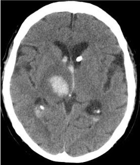
Charcot-Bouchard aneurysms are microaneurysms that are less than 1 mm in diameter. They occur in the small penetrating arterioles that perfuse the basal ganglia, pons and subcortical white matter. These aneurysms occur due to long-standing hypertension and are prone to rupture. Hemorrhagic stroke would be evident on the intial CT scan as a focus of intraparenchymal hyperdensity.

Lacunar infarcts are small ischemic infarcts ( less than 15 mm in diameter), usually involving the basal ganglia, pons, internal capsule, or deep white matter of the brain. Lacunar infarcts occur most often due to hypertensive arteriolosclerosis of small, penetrating arterioles.

21 Şubat 2014 Cuma

Neonatal complications of diabetes during pregnancy


  • Premature delivery
  • Fetal macrosomia
  • Neural tube defects (e.g Caudal regression syndrome)
  • Hypoglycemia
  • Hypocalcemia
  • Polycythemia
  • Respiratory distress
  • Transient hypertrophic cardiomyopathy.