31 Aralık 2015 Perşembe

Congenital Pyloric Stenois - Important Signs


1.  String sign: thin column of barium leaking through the tightened muscle
2. Shoulder sign: filling defect in the antrum due to prolapse of muscle inward
3.  Mushroom sign: hypertrophic pylorus against the duodenum
4. Railroad track sign: excess mucosa in the pyloric lumen resulting in 2 columns of barium.

Abnormal Pulses and Possible Etiologies

Abnormal Pulses 

Pulsus alternans: sign of left ventricular systolic dysfunction


Pulsus bigeminus: sign of hypertrophic obstructive cardiomyopathy
(HOCM)


Pulsus bisferiens: in aortic regurgitation


Pulsus tardus et parvus: aortic stenosis


Pulsus paradoxus: cardiac tamponade and tension pneumothorax


Irregularly irregular: atrial fibrillation


27 Aralık 2015 Pazar

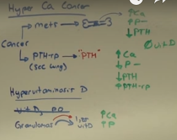
Hypercalcemia of malignancy

Hypercalcemia of malignancy

Paraneoplastic Syndromes

Cancer

Metastasis :  PTH low , High Ca , High Phosphate

(Squamous cell )PTH-rp in Lung : PTH low , High PTH r-p , High Ca , low Phosphate

4 Aralık 2015 Cuma

Amyloidosis

Epidemiology

  • Extracellular deposit of insoluble polymeric protein fibrils in tissue & organs
  • Can be primary (AL type) or secondary (AA) to chronic inflammatory conditions such as:
  • Inflammatory arthritis (e.g RA)
  • Chronic infections (e.g bronchiectasis, tuberculosis, osteomyelitis)
  • Inflammatory bowel disease (e.g Crohn's disease)
  • Malignancy (e.g. lymphoma)
  • Vasculitis

Clinical presentation

  • Asymptomatic proteinuria or nephrotic syndrome 
  • Restrictive cardiomyopathy
  • Hepatomegaly
  • Peripheral neuropathy &/or autonomic neuropathy
  • Visible organ enlargement (e.g. macroglossia)
  • Bleeding diathesis
  • Waxy thickening, easy bruising of skin

Diagnosis 

  • Tissue biopsy (e.g. abdominal fat pad)

Congo red stain shows amyloid deposits within vessel walls. 


Congo red stain shows apple green birefringence under polarized light.

List of Granulomatous diseases


  • Bartonella henselae(cat scratch disease)
  • Berylliosis
  • Churg-Strauss syndrome
  • Crohn disease
  • Francisella tularensis
  • Fungal infections (e.g., histoplasmosis, 
  • blastomycosis)
  • Granulomatosis with polyangiitis (Wegener)
  • Listeria monocytogenes(granulomatosis 
  • infantiseptica)
  • M. leprae(leprosy; Hansen disease)
  • M. tuberculosis
  • Treponema pallidum(tertiary syphilis)
  • Sarcoidosis 
  • Schistosomiasis

Th1cells secrete γ-interferon, activating macrophages. TNF-αfrom macrophages 
induce and maintain granuloma formation. Anti-TNF drugs can, as a side effect, cause 
sequestering granulomas to breakdown, leading to disseminated disease. Always test for 
latent TB before starting anti-TNF therapy.

1 Aralık 2015 Salı

Features of constrictive pericarditis


Features of constrictive pericarditis

Etiology 
  • Idiopathic or viral pericarditis
  • Cardiac surgery or radiation therapy
  • Tuberculous pericarditis (in endemic areas)


Clinical presentation

  • Fatigue and dyspnea on exertion
  • Peripheral edema and ascites
  • Increased JVP
  • Pericardial knock may be heard
  • Pulsus paradoxus
  • Kussmaul's sign


Diagnostic findings
  • ECG may be nonspecific or show atrial fibrillation or low voltage QRS complex
  • Imaging shows pericardial thickening and calcification
  • Jugular venous pulse tracing shows prominent x and y decents.

30 Kasım 2015 Pazartesi

Findings of Cor pulmonale

Characteristic findings of Cor pulmonale


Common etiologies :

  • COPD
  • Interstitial lung disease
  • Pulmonary vascular disease (eg, thromboembolic)
  • Obstructive sleep apnea


Symptoms :

  • Dyspnea on exertion, fatigue, lethargy
  • Exertional syncope (due to decreased CO)
  • Exertional angina (due to increased myocardial demand)


Examination : 

  1. Peripheral edema
  2. Increased Jugular venous pressure with prominent a wave
  3. Loud S2
  4. Right sided heave
  5. Pulsatile liver from congestion
  6. Tricuspid regurgitation murmur


Imaging :

  • EKG : partial or complete RBBB , right axis deviation, right ventricular hypertrophy right ventricular enlargement

  • Echocardiogram : Pulmonary hypertension, dilated right ventricle, tricuspid regurgitation


Right heart catheterization: Gold standard for Dx showing right ventricular dysfunction, pulmonary HTN and no left heart disease.

28 Kasım 2015 Cumartesi

Difference between Hydrocele and Varicocele

Hydrocele:--Painless enlargement of the Scrotum.
Can be Unilateral or Bilateral.

Diagnosis with Transillumination. However transillumination cannot confirm the diagnosis because it also may indicate hernia.

Ultrasound can be done to confirm the diagnosis.

Varicocele:-Varicocele is a mass of enlarged veins that develops in the spermatic cord.

A varicocele can develop in one testicle or both, but in about 85% of cases it develops in the left
testicle. The left spermatic vein drains into the renal vein .

Highest in men between the ages of 15 and 25.
The sudden appearance of varicocele in an older man
may indicate a renal tumor blocking the spermatic
vein.

S&S:--Asymptomatic or may have:--

  • Ache in the testicle
  • Feeling of heaviness
  • Infertility
  • Atrophy of the testicle
  • Visible or palpable enlarged vein 


Diagnosis:--Large varicoceles may be seen with the naked eye. Medium-sized varicoceles may be detected during physical examination by palpating--"Bag of Worms

Doppler ultrasonography uses ultrasound echos to detect the characteristic sound of the backflow of
blood through the valve.

Treatment:--Surgical Ligation of the affected Spermatic veins.

Summary :

varicocele is described as a "bag of worms", will not transilluminate
hydrocele is collection of fluid in tunica vaginalis, will feel different depending whether it is communicating or not, will transilluminate

10 Kasım 2015 Salı

Types of Miscarriage

Threatened 

  • Vaginal bleeding
  • Closed cervical os
  • Fetal cardiac activity


Missed
  • No vaginal bleeding
  • Closed cervical os
  • No fetal cardiac activity or empty sac


Inevitable

  • Vaginal bleeding
  • Dilated cervical os
  • Products of conception may be seen or felt at or above cervical os


Incomplete 

  • Vaginal bleeding
  • Dilated cervical os 
  • Some products of conception expelled and some remain


Complete

  • Vaginal bleeding or none
  • Closed cervical os 
  • Products of conception completely expelled

9 Ekim 2015 Cuma

Drugs that cause Hyperkalemia


Hyperkalemia
  • ACEI
  • ARBs
  • Beta-Blockers
  • Spironolactone
  • Other : Trimethoprim and Pentamidine 

29 Eylül 2015 Salı

Endocrine (USMLE) high yield Cases

Case: Primary psychogenic polydipsia


Case: Pheochromocytoma


Case: DKA (Diabetic ketoacidosis)


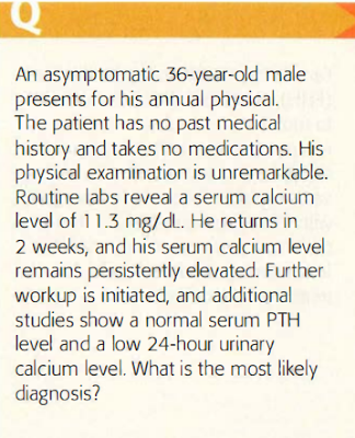
Case: Familial hypocalciuric hypercalcemia

21 Eylül 2015 Pazartesi

Warfarin and INR Testing USMLE high yield by Dr.Mirza Adnan

The Risk of Bleeding increases with an increase in the INR

Correction of excess anticoagulation is dependent upon the INR value and the presence of clinically significant bleeding


INR < 5, no significant bleeding   --> omit next warfarin dose
INR 5-9, no significant bleeding   --> stop warfarin temporarily
INR > 9  --> stop warfarin, give oral vitamin K

20 Eylül 2015 Pazar

Cardiology

Internal Medicine

USMLE Based Questions

American Diabetes Association (ADA) guidelines for Diabetes mellitus

According to the American Diabetes Association (ADA) guidelines , Diabetes Mellitus (DM) screening should start at age 45 in patients with no DM risk factors. However, patients with risk factors require screening at an earlier age.

The risk factor for developing future DM include excess body weight, family history of type 2 diabetes, and hypertension.


ADA guidelines recommend one of the following DM screening tests.

Plasma HbA1c is a screening modality for diabetes mellitus. Fasting plasma glucose, oral glucose tolerance test, and hyperglycemia in the presence of symptoms can also be used. However the results should be confirmed by repeat testing in the absence of unequivocal hyperglycemia.

18 Eylül 2015 Cuma

Retinoblastoma (intra-ocular tumor)

Retinoblastoma is the most common intra-ocular tumor of childhood. The underlying pathology involves inactivation of the Rb suppressor gene, which may be familial or sporadic.
It is highly malignant tumor, and failure to diagnose and treat the disease early may lead to death from liver and brain metastases. The other manifestations of the disease may include strabismus, decreased vision, ocular inflammation, eye pain, glaucoma, and orbital cellulitis. The diagnosis is highly suspected with Ultrasound or CT scan findings of a mass with calcifications.

Tumor suppressor gene : Rb
Associated tumor : Retinoblastoma , Osteosarcoma
Gene product : Inhibits E2F; blocks G1 -> S phase.

Diagnostic findings : 

Circular grouping of dark tumor cells surrounding pale neurofibrils
Homer-Wright rosettes (neuroblastoma, medulloblastoma, retinoblastoma)

MEDICAL MNEMONICS FOR USMLE, PLAB, AMC preparation

Common Causes of Anion gap metabolic acidosis

Common Causes of Anion gap metabolic acidosis:

1.Lactic acidosis: Hypoxia, poor tissue perfusion, mitochondrial dysfunction

2. Ketoacidosis: Type I diabetes, starvation or alcoholism

3. Methanol ingestion : Formic acid accumulation

4. Ethylene glycol ingestion : Glycolic and oxalic acid accumulation

5. Salicylate poisoning: Causes concomitant respiratory alkalosis

6. Uremia (ESRD): Failure to excrete H+ as NH4+

17 Eylül 2015 Perşembe

Indications for Dialysis

INDICATIONS FOR URGENT DIALYSIS

Remember : 

AEIOU

A Acid base disorders : Acidemia

E Electrolyte disturbance : Hyperkalemia

I Intoxication i.e methanol, ethylene glycol, lithium , salicylates

O Overload of Volume

U Uremia : Pericarditis , encephalopathy

15 Eylül 2015 Salı

Retroperitoneal hematoma following femoral arterial catheterization

Cardiac catheterization is typically done by cannulating the femoral artery to access the cardiac vessels. A common complication is hematoma formation in the soft tissue of the upper thigh. If the initial arterial puncture was done above the inguinal ligament, this hematoma can extend directly into the retroperitoneal space and cause significant bleeding, with hypotension and tachycardia.
Patient can also develop ipsilateral flank pain/back pain and neurologic deficits on the ipsilateral side.

The next step in management is to obtain a CT scan of the abdomen and pelvis without contrast to confirm the diagnosis. Treatment is mainly supportive (e.g, blood transfusion, intravenous fluids, and bed rest), with intensive monitoring. If the bleeding continues or the patient is hemodynamically unstable, the patient might need systemic reversal of anticoagulation. Patients who develop neurologic deficits in the ipsilateral extremity require urgent decompression of the hematoma.

Retroperitoneal hemorrhage from an extension of a local vascular hematoma is a common iatrogenic complication of cardiac catheterization. Diagnosis can be confirmed with abdominal CT scan, and treatment is largely supportive.

13 Eylül 2015 Pazar

Legg-Calve-Pethes disease


Legg-Calve-Pethes disease is characterized by osteonecrosis of the femoral head. It typically present in boys age 4-10 years with insidious-onset hip or knee pain and an antalfic gait.

11 Eylül 2015 Cuma

Osler Weber Rendu Syndrome

Osler Weber Rendu Syndrome :

Patients with hereditary telengiectasia can develop pulmonary AVMs associated with hemoptysis and right to left shunt physiology. Also with recurrent nose bleeds and oral lesions.

11 Ağustos 2015 Salı

Staging of Cervical Cancer

Staging of Cervical cancer.


Anatomic display of  the stages of  cervix cancer, defined by location, extent of tumor, frequency of  presentation, and  5-year survival

Causes of Vaginitis - USMLE high yield Step 2 CK

5 Ağustos 2015 Çarşamba

Placental implantation


Placental implantation. (A)  Normal placenta.  (B)  Low implantation.  (C)  Partial placenta  previa.  (D)  Complete placenta  previa.

3 Ağustos 2015 Pazartesi

Complications of Pregestational Diabetes Mellitus

OBSTETRIC ANALGESIA AND ANESTHESIA

Uterine contractions and  cervical dilation  result in visceral pain (T10-L1).
Descent of  the  fetal  head and pressure on  the  vagina and perineum  result in somatic pain (pudendal nerve, S2-S4).
In the  absence  of a  medical  contraindication,  maternal  request  is  a  sufficient medical indication for pain relief  during labor.

Absolute  contraindications  to  regional  anesthesia  (epidural,  spinal,  or 
combination) include the following: 


  • Refractory maternal hypotension 
  • Maternal coagulopathy 
  • Maternal  use  of  a  once-daily  close  of  low-molecular-weight heparin within  12  hours 
  • Untreated  maternal bacteremia 
  • Skin infection over the site  of  needle placement 
  • Increase in  ICP caused by a mass lesion

2 Ağustos 2015 Pazar

Pneumococcal polysaccharide vaccine (PPSV) for Adults

Pneumococcal polysaccharide vaccine (PPSV) should be given once to all adults age > 65. it should also be given to all adults age < 65 with chronic diseases:


Chronic lung disease

  • Asthma
  • COPD
  • Emphysema


  • Chronic cardiovascular disease
  • Diabetes mellitus
  • Chronic liver disease
  • Alcoholism
  • Cochlear implants
  • CSF leaks
  • Immunocompromising diseases

Functional or anatomic asplenia

  • Sickle cell
  • Other hemoglobinopathies
  • Splenectomy
  • Acquired asplenia.

27 Temmuz 2015 Pazartesi

WEEKS PRENATAL DIAGNOSTIC TESTING

Prenatal Diagnostic Testing Schedule

WEEKS  PRENATAL  DIAGNOSTIC TESTING

Prenatal visits  

  • Weeks 0-28: Every 4 weeks. 
  • Weeks 29-35: Every 2 weeks. 
  • Weeks 36-birth: Every week. 

Initial visit :

Heme:  CBC, Rh factor ,  type  and  screen.

Infectious disease:

UA and culture,  rubella  antibody titer , HBsAg,
RPR/VDRL, cervical gonorrhea and chlamydia, PPD, HIV,  Pap
smear (to check for dysplasia). Consider HCV and varicella based
on history.

If indicated: HbA, sickle cell screening.
Discuss genetic screening:  Tay-Sachs disease, cystic fibrosis.

9- 14  weeks :

Offer PAPP-A +  nuchal transparency + free  B-hCG +/- chorionic
villus sampling  (CVS)

15-22 weeks :

Offer maternal  serum a-fetoprotein  (MSAFP)  or quad screen  (AFP,
estriol,  P-hCG, and inhibin A)  +/- amniocentesis.

18-20 weeks :

Ultrasound for full  anatomic screen.

24-28 weeks : 

One-hour glucose  challenge test for gestational diabetes screen.

28-30 weeks :

RhoGAM for  Rh-8 women (after antibody screen).

35-40 weeks :

Group  B  strep  culture  (GBS);  repeat CBC.

34-40 weeks :

Cervical chlamydia  and  gonorrhea cultu res,  HIV,  RPR  in high-risk patients.

14 Temmuz 2015 Salı

Common Antibodies and Their Disease Associations

Common Antibodies and Their Disease Associations

ANTIBODY  DISEASE ASSOCIATION


  1. ANA  SLE 
  2. Anti-CCP  RA 
  3. Anti  centromere  CREST  syndrome 
  4. Anti-dsDNA  SLE 
  5. Anti  histone  Drug-induced  SLE 
  6. Anti-Jo- 1  Polymyositis/dermatomyositis 
  7. Anti  mitochondrial  Primary biliary cirrhosis 
  8. Antinuclear  Scleroderma 
  9. Anti-Scl-70  Scleroderma 
  10. Anti-Sm  SLE 
  11. Anti-smooth muscle  Autoimmune hepatitis 
  12. Antitopoisomerase I  Scleroderma 
  13. Anti-TSHR  Graves'  disease 
  14. c-ANCA  Vasculitis, especially Wegener's 
  15. p-ANCA  Vasculitis,  microscopic polyangiitis 
  16. Rheumatoid factor  RA 
  17. Ul  RNP antibody  Mixed connective tissue disease

10 Temmuz 2015 Cuma

Common AIDS-defining illnesses and Opportunistic infections

Common AIDS-defining illnesses:


  • Esophageal candidiasis 
  • CMV retinitis 
  • Kaposi's sarcoma 
  • CNS  lymphoma, toxoplasmosis, PML 
  • P jiroveci pneumonia  or  recurrent bacterial  pneumonia 
  • HIV encephalopathy 
  • Disseminated  mycobacterial or fungal infection 
  • Invasive cervical cancer